
Jump Cut: X-ray video & XROMM animation
Biomechanics of male and female ACL-intact and
ACL-reconstructed athletes during a jump-cut maneuver
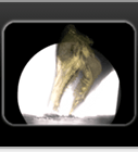
Here is an example of the data collected using X-ray Reconstruction of Moving Morphology (XROMM) for the jump-cut study. The subject's femur and tibia are shown here entering the field of view, landing, and then sidestep cutting out of the field of view. The orange bone models are shown overlaid on both X-ray sequences to illustrate the markerless tracking results obtained from Autoscoper. Take notice of the optical motion capture markers on the skin. Notice how much motion there is after impact compared to the underlying bones. We quantified how this soft tissue motion affected the interpretation of knee joint rotations and translations during the jump-cut maneuver (read more about the soft tissue artifact study).
View more about the Jump Cut and Soft Tissue Artifact studies.