
Pig Feeding
Marker-based XROMM analysis of mastication in minipigs
- Touro University College of Medicine, Hackensack, NJ
- Brown University, Providence, RI
- University of Washington, Seattle, WA
Published abstract (PDF,65KB).
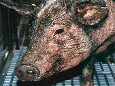 In this study we are using marker-based X-ray Reconstruction of Moving Morphology (XROMM) to visualize and measure lower jaw movements (relative to the skull) during mastication in miniature swine (Sinclair strain). The resulting XROMM animations make it possible to quantify jaw movements in 3D, study articular surface interactions in the temporomandibular joint, and visualize tooth occlusion.
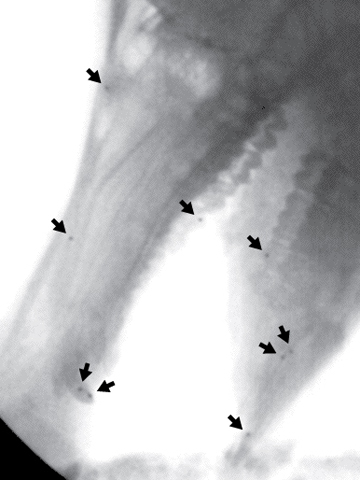 We implanted 1-mm diameter tantalum beads into the buccal aspects of several teeth, into the nasal and frontal bones, and into the body of the mandible. At least three markers are required to track each bone of interest. In two individual pigs, four markers were implanted in the mandible and five in the skull. In a third individual, five mandibular and five skull markers were implanted.
We recorded biplanar videofluoroscopy (X-Ray Movie: Lateral view | X-Ray Movie: Ventro-dorsal view) of the three little pigs feeding on pig chow and on hard nuts (in the shell). We used C-arm fluoroscopes with 12" image intensifiers, retrofitted with Photron 1024PCI cameras recording at 250 frames per second. Custom-written MATLAB scripts were used to correct image distortion, calibrate the 3D space, and extract 3D coordinate data for the radiopaque markers. Mean accuracy of marker tracking for this study was
Computed tomography (CT) scans of the three individual pigs, with tantalum beads in place, were collected and 3D models (Rotating 3D Models) reconstructed from the slices.
Autodesk Maya animation software was used to create accurate XROMM animations of fourteen mastication sequences for a total of more than 25 chewing cycles per individual pig. The motion capture data from the x-ray videos was used to animate the 3D models from CT scans. The result is a highly accurate reconstruction (XROMM Movie: Lateral view | XROMM Movie: Ventro-dorsal view) of both bone morphology and movement.
Results to date of the XROMM analysis of minipig mastication are consistent with previous studies (e.g. Herring, 1976), including bilateral grinding and frequent reversal of grinding direction with each stroke. Tracking a point on the lower incisor yields "chew loops" ("Chew Loops" Movie) similar to those observed from 2D external video.
 The 3D rigid body kinematics of the mandible confirm the importance of lateral grinding, and show for the first time that grinding results primarily from rotation of the mandible about a dorsoventrally oriented axis, with little contribution from lateral translation of the whole jaw.
 Substantial rostrocaudal translations were found, resulting from jaw protrusion and retrusion. Rotation about a rostrocaudally-oriented axis was negligible, likely due to soft tissue constraints at the TMJ.