Jump Cut

Biomechanics of male and female ACL-intact and ACL-reconstructed athletes during a jump-cut maneuver

D.L. Miranda1,2, P.D. Fadale1, M.J. Hulstyn1, R.M. Shalvoy1, J.T. Machan1,3,4,
and B.C. Fleming*1,2,5

*Author for correspondence: Braden C. Fleming | Published article

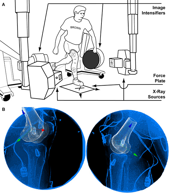
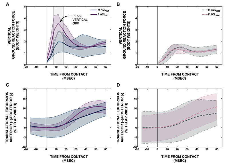
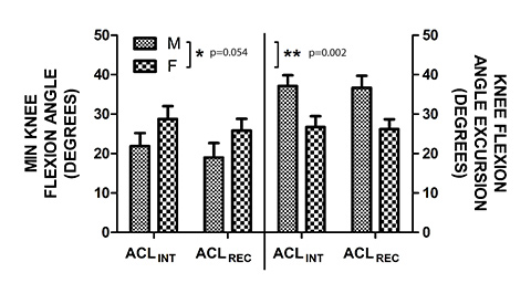
In this study we used X-ray Reconstruction of Moving Morphology (XROMM) to compare the kinetic and knee kinematic measurements from male and female ACL-intact (ACLINT) and ACL-reconstructed (ACLREC) subjects during a jump-cut maneuver. We recruited twenty subjects, 10 were ACLINT (5 males, 5 females) and 10 were ACLREC (4 males, 6 females; five years post-surgery). Each subject performed was asked to perform a jump-cut maneuver by landing on a single leg and performing a 45° side-step cut (Figure 1A). We measured the ground reaction force using a force plate and expressed the results relative to body weight. The six-degree-of-freedom knee kinematics were determined using XROMM by processing the data with Autoscoper (Figure 1B). We also used a traditional optical motion capture system to measure sagittal plane knee kinematics. We found that the ACLINT female subjects landed with a larger peak vertical GRF (p<0.001) compared to ACLINT male subjects (Figure 2A). We also observed that the ACLINT subjects landed with a larger peak vertical GRF (p≤0.036) compared to ACLREC subjects (Figure 2B). Regardless of ACL reconstruction status, we found that the female subjects underwent less knee flexion angle excursion (p=0.002) (Figure 3) and had an increased average rate of anterior tibial translation (0.05±0.01%/millisecond; p=0.037) (Figure 2C) after contact compared to the male subjects. Furthermore, the ACLREC subjects were observed to have a lower rate of anterior tibial translation compared to ACLINT subjects (0.05±0.01%/millisecond; p=0.035) (Figure 2D). Finally, no striking differences were observed in other knee motion parameters. In conclusion, this study suggests that women permit a smaller amount of knee flexion angle excursion during a jump-cut maneuver, which resulted in a larger peak vertical GRF and increased rate of anterior tibial translation. Notably, the ACLREC subjects also performed the jump cut maneuver with lower GRF than ACLINT subjects even after five years of rehabilitation. This study proposes a causal sequence whereby increased landing stiffness (larger peak vertical GRF combined with less knee flexion angle excursion) leads to an increased rate of anterior tibial translation while performing a jump-cut maneuver.

Pictures

Figure 1: A, illustration depicting the experimental set-up used to capture both biplanar videoradiography and OMC data during a jump-cut maneuver. B, example frame from the Autoscoper markerless tracking software. Each view represents one frame from each of the two videoradiographs generated from the two image intensifiers (Figure 1A). The knee shown in this image is from one of the ACL-reconstructed subjects. Both interference screws are visible in the femur and tibia. Figure 2: A, ACL-intact vertical GRF. B, ACL-reconstructed vertical GRF. C, ACL-intact AN/PO translational excursion. D, ACL-reconstructed AN/PO translational excursion. Figure 3: Left y-axis, minimum knee flexion angle for ACL-intact and ACL-reconstructed male and female subjects. Minimum flexion occurred at or immediately following ground contact. Right y-axis, knee flexion angle excursion for ACL-intact and ACL-reconstructed male and female subjects. Knee flexion angle excursion was defined as the change in knee flexion angle from minimum flexion to maximum flexion.

Movie

Jump Cut: X-ray video & XROMM animation

Reference

Miranda, D.L., P.D. Fadale, M.J. Hulstyn, R.M. Shalvoy, J.T. Machan, and B.C. Fleming. (2012). Knee Biomechanics during a Jump-Cut Maneuver: Effects of Gender and ACL Surgery. Medicine and Science in Sports and Exercise. [Epub ahead of print].
Published article.

Related Publications

Miranda, D.L., M.J. Rainbow, J.J. Crisco, and B.C. Fleming. (2013). Kinematic differences between optical motion capture and biplanar videoradiography during a jump-cut maneuver. Journal of Biomechanics. 46(3): 567-573. Published article.

Miranda, D.L., J.B. Schwartz, A.C. Loomis, E.L. Brainerd, B.C. Fleming, and J.J. Crisco. (2011). Static and dynamic error of a biplanar videoradiography system using marker-based and markerless tracking techniques. Journal of Biomechanical Engineering. 133(12): 121002. Published article.

Miranda, D.L., M.J. Rainbow, E.L. Leventhal, J.J. Crisco, and B.C. Fleming. (2010). Automatic determination of anatomical coordinate systems for three-dimensional bone models of the isolated human knee. Journal of Biomechanics. 43(8): 1623-1626.
Published article.

Author Affiliations

1Department of Orthopaedics, The Warren Alpert Medical School, Brown University
and Rhode Island Hospital, Providence, RI, USA

2Center for Biomedical Engineering, Brown University, Providence, RI, USA

3Department of Surgery, The Warren Alpert Medical School, Brown University,
Providence, RI, USA

4Research, Biostatistics, Rhode Island Hospital, Providence, RI, USA

5School of Engineering, Brown University, Providence, RI, USA