History
Below is a brief history of methods for measuring skeletal kinematics, including a definition of XROMM and some perspective on how XROMM fits in with other rapidly developing methods. At Brown University, Professors Beth Brainerd, Steve Gatesy and collaborators coined the term XROMM and began hardware and software development in 2006, based on Scientific Rotoscoping work that Gatesy had been doing since the 1990s.
XROMM development owes a great debt to previous x-ray motion analysis work, and we hope that the references below give some sense of this recent but rich history. We apologize to researchers whose contributions have been accidentally omitted, and would be happy to have them brought to our attention.
The text below is excerpted, with some modification, from: Brainerd, E.L., S.M. Gatesy, D.B. Baier, T.L. Hedrick, K.A. Metzger, J. Crisco, and S.L. Gilbert (2010). X-ray Reconstruction of Moving Morphology (XROMM): applications and accuracy in comparative biomechanics research.
Prior to 2010, most estimates of skeletal kinematics came from motion capture of external markers attached to skin or tight clothing, but these estimates suffer from poor fidelity of skin movement to underlying bone movement (Filipe et al., 2006; Lanovaz et al., 2004; Leardini et al., 2005). Furthermore, many bones and joints are simply too deeply embedded in the body for external markers to track even their gross movements.
Measuring skeletal kinematics with high accuracy also requires three-dimensional (3D) analysis. Few movements are truly planar, and even when approximately planar, the movements must be performed parallel to the film plane for two-dimensional (2D) analysis to be valid. These conditions are rarely met with sufficient accuracy for measuring the full range of translations and rotations that occur during natural joint movements.
X-ray motion analysis solves the problem of external marker movement artifacts, but to date, most x-ray studies have been limited to 2D motion analysis. Many valuable insights into animal movement and musculoskeletal biomechanics have been gained from 2D and 3D external motion analysis and 2D x-ray motion analysis, but new x-ray technologies and computational methods are now enabling true 3D x-ray motion analysis (e.g. Bey et al., 2006; Gatesy and Alenghat, 1999; Gatesy et al., 2010; Tashman et al., 1995).
We offer a new term "X-ray Reconstruction of Moving Morphology (XROMM)" to refer to a set of emerging 3D x-ray motion analysis methods that combine skeletal movement data from in vivo x-ray videos with skeletal morphology from 3D bone scans (e.g. computed tomography (CT) or laser scans). Scientific Rotoscoping is one type of XROMM analysis, in which multi-bone, articulated models are manually aligned (registered) to x-ray and standard video of animal movement (Gatesy and Alenghat, 1999; Gatesy et al., 2010). More automated methods for registering markerless bone models to biplanar x-ray movies are also emerging (Bey et al., 2008; Bey et al., 2006; You et al., 2001), with great promise for increasing the throughput of markerless XROMM analysis. These methods for markerless autoregistration build on previous work registering the movement of knee replacement devices in human patients to single plane x-ray movies (Banks and Hodge, 1996; Zuffi et al., 1999).
Implantation of radio-opaque bone markers allows bone models to be animated directly from bone marker coordinates (Tashman and Anderst, 2003). Marker-based XROMM, relative to Scientific Rotoscoping and other markerless methods, offers the possibility of higher throughput and (usually) greater accuracy (Brainerd et al., 2010; Tashman and Anderst, 2003; Tashman et al., 1995). Marker-based XROMM is also known as Dynamic Roentgen Stereophotogrammetric Analysis, particularly in the field of orthopedic biomechanics (Papaioannou et al., 2009; Tashman et al., 2007).
In the future, it is likely that the most effective application of XROMM for comparative biomechanics research will include implanting markers in the larger, more surgically accessible bones of interest, and then using markerless Scientific Rotoscoping or autoregistration for the remaining elements. The biplanar videofluoroscopy, distortion correction, calibration, and Matlab and Maya methods that we have developed lend themselves well to this hybrid approach. Biplanar videofluoroscopy, combined with our calibration and distortion correction methods, show promise for increasing the efficiency of Scientific Rotoscoping well beyond the original implementation of this method with single-plane x-ray and standard camera views (Gatesy et al., 2010). The articulated skeletal model (digital marionette) method will continue to be a valuable approach, and may be enhanced by highly accurate, marker-based animation of the proximal bones. These accurate proximal bone positions can then help guide the markerless placement of the more distal elements with Scientific Rotoscoping.
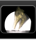
The power of XROMM analysis far exceeds the relatively simple goal of tracking specific points on the skeleton with high accuracy (Gatesy et al., 2010). The key to this greater power is the Morphology in X-ray Reconstruction of Moving Morphology. The use of high-resolution 3D bone models in XROMM means that motion can be studied in the context of detailed skeletal morphology. For example, in our work on pig mastication, the XROMM animation contains information on tooth shape as well as jaw movement, making it possible to visualize and measure the interactions of specific upper and lower tooth cusps during occlusion. Another example of this power could be the development of a new approach to the study of form-function relationships in joints. XROMM makes it possible to visualize and measure the interactions of articular surfaces within joints (Anderst and Tashman, 2003), perhaps opening up "dynamic arthrology" as a new, morphology-focused approach to the study of joint biomechanics.
References
Anderst, W.J. and S. Tashman. (2003). A method to estimate in vivo dynamic articular surface interaction. J. Biomech. 36, 1291-1299.
Banks, S.A. and W.A. Hodge. (1996). Accurate measurement of three dimensional knee replacement kinematics using single-plane fluoroscopy. IEEE Trans. Biomed. Eng. 43, 638-648.
Bey, M.J., S.K. Kline, R. Zauel, T.R. Lock, and P.A. Kolowich. (2008). Measuring dynamic in-vivo glenohumeral joint kinematics: Technique and preliminary results. J. Biomech. 41, 711-714.
Bey, M.J., R. Zauel, S.K. Brock, and S. Tashman. (2006). Validation of a new model-based tracking technique for measuring three-dimensional, in vivo glenohumeral joint kinematics. J. Biomech. Eng. 128, 604-609.
Brainerd E.L., Baier D.B., Gatesy S.M., Hedrick T.L., Metzger K.A., Gilbert S.L., Crisco J.J. (2010). X-ray reconstruction of moving morphology (XROMM): precision, accuracy and applications in comparative biomechanics research. J. Exp. Zool. 313A:262–279.
Filipe, V.M., J.E. Pereira, L.M. Costa, A.C. Maurício, P.A. Couto, P. Melo-Pinto, and A.S.P. Varejão. (2006). Effect of skin movement on the analysis of hindlimb kinematics during treadmill locomotion in rats. J. Neurosci. Methods. 155, 55–61.
Gatesy, S.M. and T. Alenghat. (1999). A 3-D computer-animated analysis of pigeon wing movement. Amer. Zool. 39, 104A.
Gatesy S.M., Baier D.B., Jenkins F.A., Dial K.P. (2010). Scientific rotoscoping: a morphology-based method of 3-D motion analysis and visualization. J. Exp. Zool. 313A:244–261.
Lanovaz, J.L., S. Khumsap, and H.M. Clayton. (2004). Quantification of three-dimensional skin displacement artefacts on the equine tibia and third metatarsus. Equine and Comparative Exercise Physiology. 1, 141-150.
Leardini, A., L. Chiari, U. Della Croce, and A. Cappozzo. (2005). Human movement analysis using stereophotogrammetry part 3. Soft tissue artifact assessment and compensation. Gait Posture. 21, 212-225.
Papaioannou, G., C. Mitrogiannis, G. Nianios, and G. Fiedler. (2009). Assessing residual bone-stump-skin-socket interface kinematics of above knee amputees with high accuracy biplane dynamic roentgen stereogrammetric analysis. 55th Annual Meeting of the Orthopaedic Research Society 2009, Paper No. 346.
Tashman, S. and W. Anderst. (2003). In vivo measurement of dynamic joint motion using high-speed radiography and CT: Application to canine ACL deficiency. J. Biomech. Eng. 125, 238-245.
Tashman, S., K. DuPre, H. Goitz, T. Lock, P. Kolowich, and M. Flynn. (1995). A digital radiographic system for determining 3D joint kinematics during movement. In 19th Ann. Meeting Amer. Soc. Biomech., pp. 249-250. Stanford, CA: ASB Press.
Tashman, S., P. Kolowich, D. Collon, K. Anderson, and W. Anderst. (2007). Dynamic function of the ACL-reconstructed knee during running. Clinical Orthopaedics and Related Research. 454, 66-73.
You, B.-M., P. Siy, W. Anderst, and S. Tashman. (2001). In vivo measurement of 3-D skeletal kinematics from sequences of biplane radiographs: Application to knee kinematics. IEEE Trans. Med. Imag. 20, 514-525.
Zuffi, S., A. Leardini, F. Catani, S. Fantozzi, and A. Cappello. (1999). A model-based method for the reconstruction of total knee replacement kinematics. IEEE Trans. Med. Imag. 18, 981-991.


